Newsletters


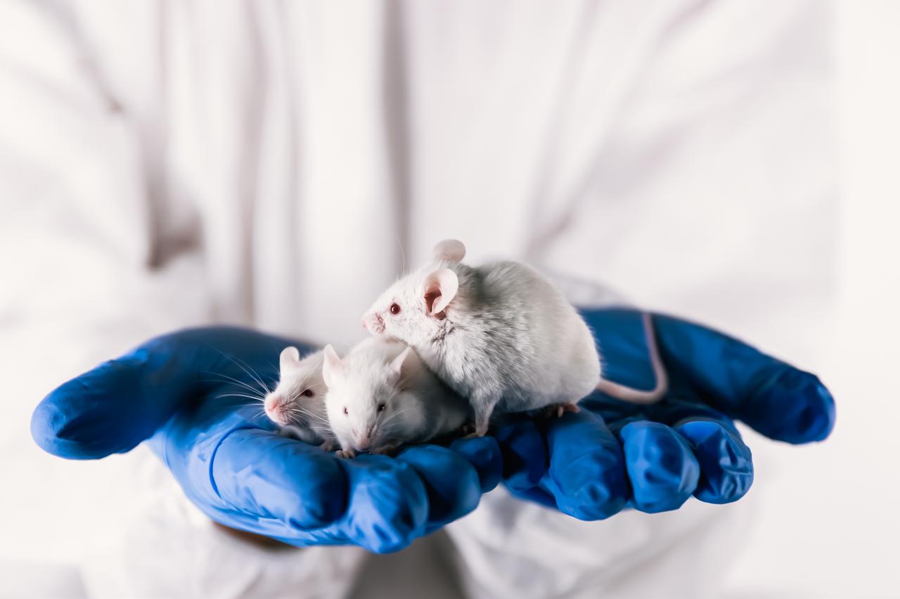


Mice in a laboratory in the US state of California move around their cages like any other mice. Their brains, however, contain millions of functioning human neurons.
Researchers at Stanford University transplanted laboratory-grown human cells into mice genetically engineered to lack certain brain regions.
These include the cerebral cortex, the brain’s outer layer, which plays a key role in perception, movement, decision-making and attention.
According to a study published in the journal Nature on Sept. 16, the animals, known as xenocortical mice, could offer a new model for studying neural circuits and brain diseases such as schizophrenia, cerebral palsy and dementia.
Placing human brain tissue in an animal raises questions about how human the mice are and what the researchers found.
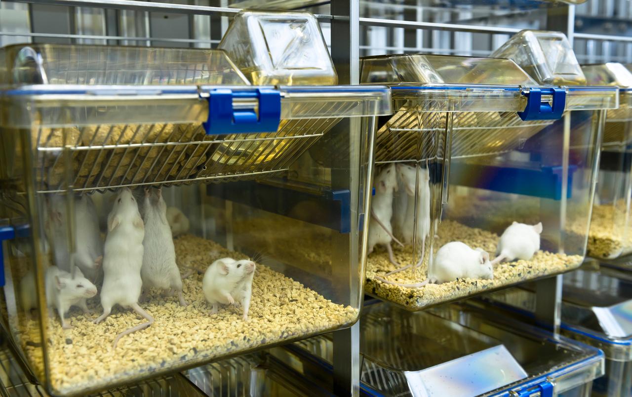

Researchers genetically modified the mice so that they were born and grew without most of the cerebral cortex and the neighboring hippocampus, a region involved in movement, learning and memory.
When the mice were two days old, researchers injected human “organoids” into the empty spaces in their brains.
Organoids are small clusters of cells grown in the laboratory from stem cells or tumor tissue that mimic real human organs. Each organoid in the experiment was grown from stem cells taken from healthy donors and contained about 100,000 human cells.
Researchers removed about 14 million neurons from the mice and added about 4 million human neurons in their place.
The researchers stressed that the procedure is not equivalent to a brain transplant and that the animals are not “mice that think like humans.”
The procedure was tested on 29 mice and succeeded in 25.
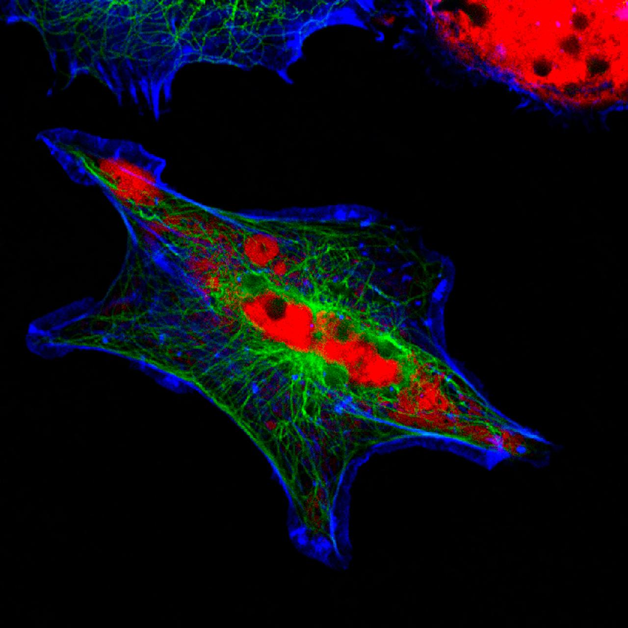
Within three months, the human tissue grew about fivefold. It connected to the animals’ blood supply and filled more than 90 percent of the cavities created in the brain.
Human tissue made up about half of the brain volume in the xenocortical mice, yet the animals walked and vocalized normally.
The researchers also ran a maze test. Mice without transplanted tissue had difficulty finding their way, while mice with human cells performed better. The result suggests the human tissue may have contributed to cognitive functions.
Another unexpected finding was that part of the transplanted tissue developed into von Economo neurons.
These long, rare nerve cells are found in humans, great apes, whales and elephants, but not normally in mice. They are considered nearly impossible to produce in a laboratory.

The study may also help research into cerebral palsy.
Human babies can be deprived of oxygen because of complications linked to a longer and more complex birth process.
Oxygen deprivation during birth is much rarer in mice, which makes it difficult to study the condition in rodents.
In the experiment, genetically modified mice exposed to low oxygen levels showed brain damage, gait changes, and impaired limb coordination.
Human brain diseases are among the most difficult health problems to study.
The human brain consists of about 86 billion nerve cells and trillions of connections between them. This biological complexity, combined with ethical and safety restrictions, makes access to living human brain tissue highly limited.
Scientists have therefore tried for years to grow organoids, or small clusters of human nerve cells, in the laboratory.
These structures are not real brains grown in a dish. They lack the connections, external input and biological environment of a living brain.
Sergiu Pasca, the study’s lead author and a professor of psychiatry and behavioral sciences at Stanford, said the method could give researchers unprecedented insight into how the human brain works.
“For the past 20 years, efforts have been made to build models of the human brain outside the human body,” Pasca said.
Pasca said the study will not replace all earlier models. “But it will provide access to different aspects of human brain function that would otherwise be extremely hard to study,” he said.


 Set as preferred source
 Set as preferred source



 2 min read