Newsletters


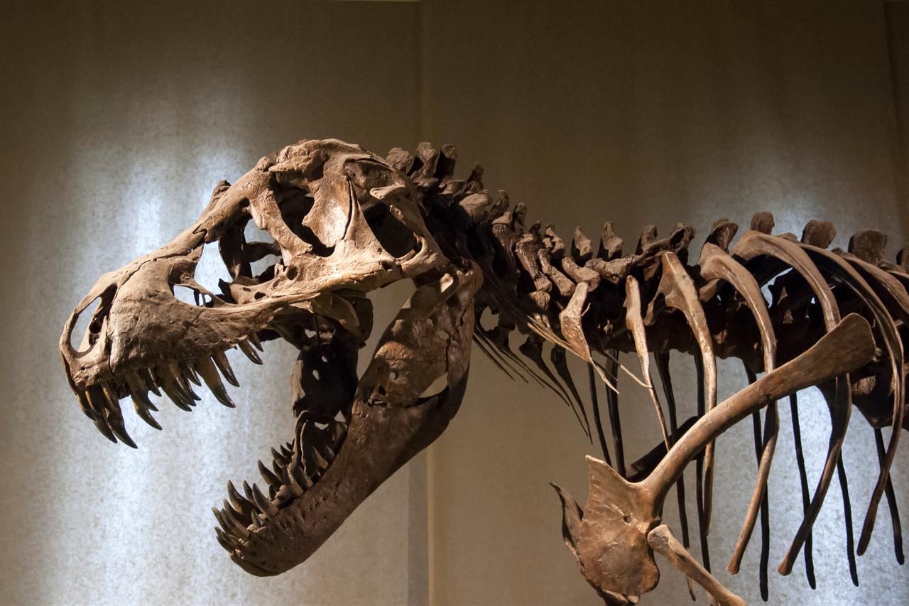


Microscopic structures and chemical traces found in a 115 million-year-old dinosaur fossil from Brazil may be preserved red blood cells, according to a new study. An independent expert disputes that interpretation.
The research was published Nov. 2 in the journal PNAS. The team reports it identified structures resembling red blood cells alongside chemical signatures of heme, the iron-containing molecule that carries oxygen in hemoglobin. The authors describe the result as the oldest chemical evidence of blood-related material in a dinosaur fossil.
The material comes from the limb bones of Aratasaurus museunacionali, an extinct theropod about 3 meters (10 feet) long that lived in Brazil during the Early Cretaceous period. Theropods were mostly meat-eating dinosaurs that walked on two legs, a group that also includes Tyrannosaurus rex and Velociraptor.
Earlier research has detected blood in dinosaur fossils. The new study is the first to use chemical analysis to identify heme-based signatures in dinosaur bone, according to its authors.
Kliti Grice, a professor in the School of Earth and Planetary Sciences at Curtin University in Australia and a study co-author, said the team was not looking for blood at the outset. Its starting point was a broader question about how much of an ancient organism’s biology survives fossilization. Grice said exceptionally preserved fossils may hold “microscopic structures and molecular traces of the original organism.”
The researchers combined microscopy, mineral analysis and molecular geochemistry. They found numerous microscopic structures inside vascular canals, channels that once carried blood vessels through the bone. The team says the shape, size and location of the structures are consistent with red blood cells.
Chemical analysis detected heme-related compounds associated with the structures. Grice said the tests were carried out in the bone itself, which allowed the team to link chemical signals to specific structures in their physical setting. She said this approach is stronger than extracting molecules from a fossil and assuming where they came from.
The fossil also contained amino acids associated with collagen, the main structural protein in bone.
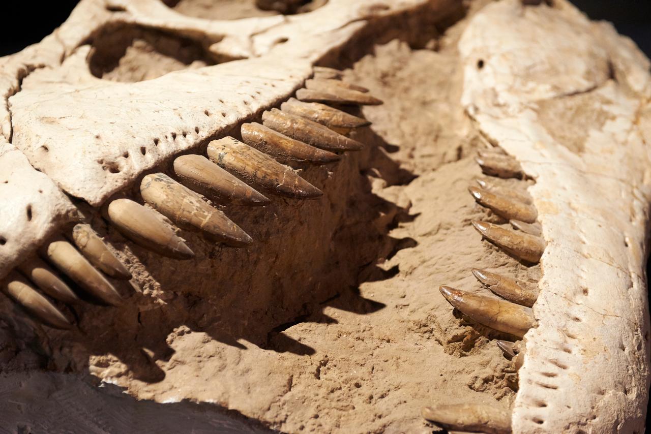

The team suggests the animal was buried quickly by sediment in a low-oxygen environment containing microbial mats, thin films made of microorganisms. Such conditions may have slowed normal decomposition and helped preserve the microscopic structures and their chemistry.
Grice said the red blood cell-like structures were mineralized with hausmannite, a manganese oxide mineral. She said early mineralization may have stabilized or replicated the structures before they were destroyed, while some of the associated chemistry survived in altered form.
Mary Schweitzer, a molecular paleontologist at North Carolina State University who was not involved in the study, said she is not convinced the images show red blood cells. Schweitzer, who has studied soft tissues from the dinosaur age, agreed that molecules produced within the body can persist over geological time and offer valuable information.
She said the structures do not convincingly match the expected appearance of red blood cells in nonmammalian animals. Mammalian red blood cells lose their nuclei during development, while those of birds and reptiles keep them, which typically gives the cell a nuclear bulge. Schweitzer said the structures in the study are more concave, show no such bulge and are smaller than most nonmammalian red blood cells, so “morphological similarity is not met.”
Schweitzer also welcomed the team’s use of new technology and said she is excited to see researchers moving toward molecular approaches to ancient fossils.


 Set as preferred source
 Set as preferred source



 1 min read